
A hole in the eardrum may be tiny, but it can crperforated eardrum can muffle hearing, trigger repeated infections, cause drainage, and make swimming feel like an extreme sport.
Fortunately, modern eardrum repair offers several ways to close the opening and rebuild the middle ear. Some repairs take only a few minutes in an office or operating room. Others involve reconstructing the eardrum, repairing the tiny hearing bones, or removing chronic disease. In straightforward cases, closure rates are commonly around 80% to 90%, although results vary according to the condition of the ear and the definition of “success.”
What Is Eardrum Repair?
The eardrum, medically known as the tympanic membrane, is a thin layer of tissue separating the ear canal from the middle ear. When sound waves reach it, the membrane vibrates and transfers that energy to three tiny bonesthe malleus, incus, and stapes. Those bones then pass the signal toward the inner ear.
A perforation prevents the membrane from vibrating normally. It can also create an open doorway through which water and microorganisms enter the middle ear. The result may be conductive hearing loss, recurring infection, discomfort, tinnitus, or drainage.
Eardrum repair refers to procedures that close the perforation and restore a stable barrier between the ear canal and middle ear. If the hearing bones are damaged, the surgeon may reconstruct them during the same operation. auses an Eardrum Perforation?
Common causes include:
- Middle ear infections that build pressure behind the eardrum
- Chronic draining ear disease
- Objects placed too deeply in the ear canal
- Sudden pressure changes from diving, flying, or an explosion
- A slap or direct injury to the ear
- Previous ear-tube placement or other ear surgery
- Cholesteatoma, an abnormal collection of skin cells in the middle ear
Many fresh, small perforations heal without an operation. Surgery becomes more likely when the opening remains for several months, causes repeated infections, interferes with hearing, or prevents safe participation in water activities.
Types of Eardrum Repair
Patients often hear the terms patching, myringoplasty, and tympanoplasty used as though they are interchangeable. They overlap, but each can describe a different level of repair.
1. Office-Based Eardrum Patching
A small, uncomplicated hole may be treated by carefully irritating or “freshening” its edges and placing a thin patch over the opening. The patch acts like temporary scaffolding, allowing new cells to grow across the gap.
Materials may include paper, gel foam, biologic material, or another specialized membrane. This approach is less invasive than formal tympanoplasty, but it is generally best suited to selected small perforations. More than one application may be required.
2. Myringoplasty
Myringoplasty repairs the eardrum without reconstructing the middle-ear bones. It may involve a patch or a graft made from the patient’s tissue. Common graft sources include fascia, the strong connective tissue covering the temple muscle, and perichondrium or cartilage harvested near the ear.
The overall closure rate for myringoplasty is often reported near 90%, although the result depends on perforation size, location, infection history, tissue quality, and surgical technique. e I Tympanoplasty
Type I tympanoplasty is a formal reconstruction of the tympanic membrane when the hearing bones remain intact. In practice, the distinction between myringoplasty and Type I tympanoplasty is sometimes fuzzy. The important point is that the operation focuses on closing the membrane rather than rebuilding the ossicular chain.
4. Tympanoplasty With Ossiculoplasty
Long-term infection, trauma, or cholesteatoma can erode or disconnect the malleus, incus, or stapes. Closing the eardrum alone would then create a healthy-looking membrane that still cannot transmit sound effectively.
Ossiculoplasty repairs or replaces damaged hearing bones using reshaped natural bone, cartilage, or a small prosthesis. This operation is more complex, and hearing results can be less predictable than the simple closure of an isolated eardrum hole. panoplasty With Mastoid or Cholesteatoma Surgery
If destructive infection or cholesteatoma extends behind the eardrum, the surgeon’s first priority is creating a safe, dry ear. Diseased tissue may need to be removed from the middle ear or mastoid bone before reconstruction begins.
This is no longer a simple patch job. It is closer to renovating the plumbing, flooring, and electrical system before hanging new wallpaper. Hearing restoration remains important, but complete disease removal and infection control come first.
6. Endoscopic and Microscopic Tympanoplasty
These terms describe how the surgeon sees the operating field. Traditional microscopic tympanoplasty uses an operating microscope and may require an incision behind or near the ear. Endoscopic ear surgery uses a narrow camera inserted through the ear canal, offering a wide view of areas that can be difficult to see with a straight-line microscope.
An endoscopic approach can sometimes avoid an external incision, but it is not automatically superior for every patient. Microscopes provide depth perception and allow two-handed surgery, while endoscopes offer close panoramic views around corners. Many surgeons use both tools when appropriate.
How Is the Graft Positioned?
Underlay Technique
With an underlay repair, the graft is placed beneath the remaining eardrum and usually under part of the malleus. It is widely used because it is reliable and technically straightforward for many central perforations.
Overlay Technique
With an overlay repair, the graft is placed over the supporting ring of the eardrum after the surface tissue has been lifted. It may be useful for large, anterior, or near-total perforations, but it requires careful positioning to avoid thickening, poor healing, or displacement.
Cartilage Versus Fascia
Temporalis fascia is thin and behaves similarly to a natural eardrum. Cartilage is stiffer and may better resist retraction or recurrent perforation in ears with poor pressure regulation, revision surgery, or large defects.
Systematic reviews suggest that cartilage can provide stronger anatomical closure than fascia in some situations, while average hearing outcomes are generally similar. The best material is therefore not a universal winner; it is the one suited to the ear being repaired.
What Happens Before Surgery?
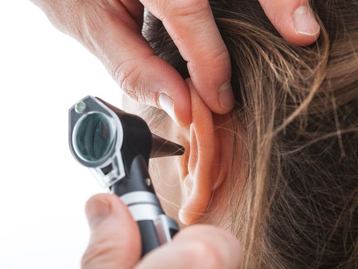
An ENT specialist examines the ear under magnification and records the perforation’s size, position, condition, and relationship to the hearing bones. The clinician also looks for active infection, scar tissue, retraction, or cholesteatoma.
A hearing test measures air and bone conduction and helps determine whether hearing loss comes only from the perforation or also involves the ossicles or inner ear. Speech testing may be included. CT imaging is not routinely necessary for a simple hole, but it may be ordered when cholesteatoma, unusual anatomy, or extensive disease is suspected. rainage may need treatment before surgery. Patients should also tell the surgical team about smoking, diabetes, blood-thinning medications, previous ear procedures, allergies, and difficulty equalizing ear pressure.
Eardrum Repair Success Rates
Published success rates vary because surgeons and researchers do not always measure the same outcome. A technically intact graft is not identical to better hearing, and better hearing is not identical to eliminating infection.
Anatomical Success
Anatomical success means the graft heals and the eardrum remains closed. Major U.S. medical centers commonly describe closure rates of approximately 75% to 90%, while broader studies report ranges from roughly 75% to 98%. Straightforward primary repairs tend to land at the higher end; revision surgery and complicated chronic ear disease may fall lower. g Success
Hearing often improves when the perforation is closed and the ossicles move normally. However, the final result depends on the inner ear, the condition of the hearing bones, scar tissue, middle-ear ventilation, and whether disease has caused permanent damage.
A perfectly healed graft cannot reverse unrelated sensorineural hearing loss. This is why postoperative audiograms matter more than enthusiastic declarations that the eardrum “looks great.” The ear deserves both a beauty inspection and a performance test.
Infection-Control Success
Even when hearing improvement is modest, closing a perforation can reduce drainage, protect the middle ear from contaminated water, and make the ear easier to manage. For someone with years of infections, a reliably dry ear can be a major quality-of-life improvement.
Factors That Influence the Result
- Perforation size: Larger or near-total holes may be more difficult to close.
- Perforation location: Anterior and marginal openings can be technically challenging.
- Middle-ear infection: Ongoing inflammation can interfere with graft healing.
- Eustachian tube function: Poor ventilation may pull the repaired membrane inward.
- Previous surgery: Scar tissue and altered anatomy can complicate revision repair.
- Cholesteatoma: Disease removal adds complexity and may require staged reconstruction.
- Smoking: Tobacco exposure can reduce blood flow and tissue healing.
- Diabetes or other health conditions: Poorly controlled disease may impair recovery.
- Postoperative care: Water exposure, pressure changes, and premature strenuous activity may threaten a fresh graft.
Research does not agree on the importance of every factor in every population, but perforation characteristics, middle-ear health, Eustachian tube function, smoking, and surgical complexity repeatedly appear in outcome studies.
Possible Complications of Eardrum Repair
Tympanoplasty is generally safe, and serious complications are uncommon. Still, surgery takes place within millimeters of the hearing and balance organs, facial nerve, and taste nerve, so the risk discussion deserves more than a cheerful shrug.
Graft Failure or Recurrent Perforation
The graft may fail to attach completely, or a new hole may develop later. A small residual opening can sometimes be patched, while a larger failure may require revision tympanoplasty.
Infection or Persistent Drainage
Mild bloody drainage can occur during early recovery. Increasing pain, pus-like discharge, foul odor, or fever may indicate infection and should be reported to the surgical team.
Temporary or Permanent Hearing Changes
Hearing usually sounds worse immediately after surgery because of swelling and packing. Permanent worsening is uncommon when repair is limited to the membrane, but the risk is greater when the ossicles or extensive middle-ear disease must be treated.
Taste Disturbance
The chorda tympani nerve, which carries taste information from part of the tongue, crosses the middle ear. Stretching or disturbing it can cause a metallic taste, altered flavor, or tongue numbness. Symptoms often improve over weeks or months.
Dizziness, Tinnitus, or Balance Problems
Brief dizziness or increased ringing may occur after surgery. Severe, persistent, or worsening vertigo deserves prompt evaluation.
Facial Nerve Injury
Facial weakness is rare but serious. New asymmetry, difficulty closing an eye, or weakness around the mouth requires immediate contact with the surgeon.
Anesthesia and Incision Risks
General surgical risks include bleeding, medication reactions, infection, scarring, and numbness around an incision. Individual risk depends on health status and the extent of surgery.
Recovery After Tympanoplasty
Most patients return home on the day of surgery. Ear pressure, mild pain, popping, crackling, and light bloody drainage can occur during the first several days. Because packing blocks the canal, hearing may seem impressively terrible before it gets better. That is usually part of the process rather than the eardrum filing a formal complaint.
Routine discomfort often improves within 10 days to two weeks. The graft itself heals much more slowly, and hearing may continue changing for two or three months. Follow-up examinations and a postoperative hearing test are used to confirm both closure and functional improvement. Recovery Instructions
- Keep the operated ear dry until the surgeon permits normal water exposure.
- Do not remove packing unless specifically instructed.
- Avoid swimming and submerging the ear.
- Do not blow the nose forcefully during early healing.
- Sneeze with the mouth open to reduce pressure in the middle ear.
- Avoid heavy lifting, straining, and impact exercise until cleared.
- Ask the surgeon when flying or diving will be safe.
- Use prescribed drops and medications exactly as directed.
When to Call the Surgeon
Contact the surgical team for heavy bleeding, increasing pain, high fever, foul-smelling discharge, persistent vomiting, severe dizziness, sudden hearing deterioration, worsening tinnitus, or facial weakness.
Practical Experiences: What Eardrum Repair Often Feels Like
The following scenarios combine commonly reported recovery patterns for educational purposes. They are not testimonials from specific patients, and individual experiences can differ significantly.
The “Why Is My Hearing Worse?” Phase
One of the most surprising experiences after eardrum repair is temporary hearing reduction. A patient may arrive expecting an instant audio upgrade and leave feeling as though someone placed a pillow over the operated ear.
This usually happens because the ear canal contains packing and the middle ear is swollen. Fluid, dried blood, ointment, and dissolvable material can also block sound. Crackling and popping may appear as the packing shifts or begins to dissolve. Hearing often changes from day to day, so judging the final result during the first week is like reviewing a cake while it is still batter.
The Recovery That Is Not a Straight Line
Patients often feel significantly better after several days and then notice temporary fullness, mild discomfort, or strange sounds again. Normal recovery can fluctuate. A mildly frustrating day does not automatically mean the graft has failed.
What matters is the overall direction and the presence of warning signs. Increasing pain, heavy drainage, severe vertigo, or fever is different from occasional pressure or popping. A written list of symptoms and dates can help the surgeon determine whether the course is normal.
The Water-Avoidance Challenge
Keeping an ear dry sounds easy until shower day. Patients commonly find that a petroleum-jelly-coated cotton ball at the canal entrance provides a practical splash barrier when approved by the surgeon. It should not be pushed deeply into the ear.
Parents may need extra creativity with young children who consider bathwater a competitive sport. Calm supervision, shallow baths, and delaying swimming until medical clearance can protect the graft during its most vulnerable period.
The First Follow-Up Visit
The initial follow-up may involve inspecting the incision, removing or adjusting packing, and checking for infection. The eardrum can still look thick, swollen, or dull. Patients sometimes expect the surgeon to announce a final success immediately, but complete healing cannot always be confirmed that early.
Later visits are often more informative. Once swelling and packing have cleared, the surgeon can evaluate the graft’s position and integrity. An audiogram then shows whether sound conduction has improved. These appointments are essential even when the ear feels fine.
Experience With Children
Children may recover physically faster than adults but can have difficulty following pressure and activity restrictions. A child who feels normal may immediately want to run, wrestle, jump on furniture, and test whether gravity has been repealed.
Parents often benefit from planning quiet activities before surgery, informing the school about temporary restrictions, and keeping medication and follow-up instructions in one visible place. Because childhood Eustachian tube function and recurring infections can affect outcomes, long-term monitoring may be particularly important.
The Emotional Side of Waiting
The toughest part for some patients is uncertainty. The operation may be over in a few hours, but meaningful hearing improvement can take months. Every pop, buzz, or muffled conversation may trigger worry.
It helps to know the surgeon’s expected recovery milestones before the procedure: when packing will be removed, when work or school can resume, when water is allowed, and when hearing will be tested. Clear expectations cannot accelerate tissue healing, but they can prevent the imagination from turning every crackle into a medical miniseries.
Questions to Ask Before Eardrum Repair
- Is observation or office patching still reasonable for my perforation?
- Will the procedure repair only the eardrum or also the hearing bones?
- Do you recommend fascia, cartilage, or another graft material?
- Will the surgery be endoscopic, microscopic, or a combination?
- What closure rate do you achieve in ears similar to mine?
- How much hearing improvement is realistically expected?
- Is there evidence of cholesteatoma or chronic mastoid disease?
- How long must I avoid water, exercise, flying, and nose blowing?
- What symptoms should trigger an urgent call?
Conclusion
Eardrum repair is not one single operation. It ranges from a simple patch for a small perforation to complex tympanoplasty with ossicular reconstruction and removal of chronic ear disease.
For uncomplicated repairs, successful closure is commonly achieved in approximately 80% to 90% of cases, with some studies reporting even higher rates. Nevertheless, anatomical closure, hearing improvement, and infection control are separate measures. The best estimate for an individual patient comes from the perforation’s size and location, middle-ear condition, hearing test, medical history, and planned surgical technique.
The operation is usually safe, but graft failure, infection, altered taste, dizziness, tinnitus, and hearing changes can occur. Careful postoperative protectionand a little patience while the ear performs its slow-motion rebuilding projectcan help the repair reach its best possible outcome.