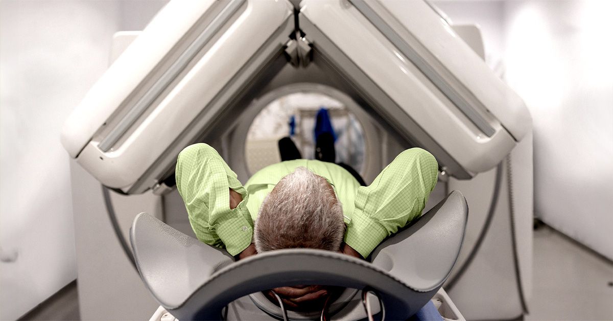

A PET scan can reveal where non-Hodgkin lymphoma is active, how widely it has spread, and whether treatment appears to be working. That makes it one of the most useful imaging tools in modern lymphoma care. Still, it is not a magical cancer detector, and it cannot replace the laboratory analysis of an actual tissue sample.
Most lymphoma imaging today is performed as a combined PET/CT scan. The PET portion highlights unusual metabolic activity, while the CT portion shows the precise location, shape, and size of lymph nodes, organs, and other structures. One scan supplies the biological clues; the other provides the anatomical map. Think of it as pairing a heat-sensing camera with a detailed road atlasminus the person in the passenger seat repeatedly saying, “You missed the turn.”
This guide explains how doctors use a PET scan for non-Hodgkin lymphoma diagnosis and staging, what the results can and cannot reveal, how to prepare, and what patients commonly experience during the process.
What Is a PET/CT Scan?
PET stands for positron emission tomography. For most lymphoma examinations, a healthcare professional injects a small amount of a radioactive tracer called fluorine-18 fluorodeoxyglucose, commonly shortened to FDG. FDG resembles glucose, the sugar cells use for energy.
Many lymphoma cells consume more glucose than surrounding normal tissue. Areas that absorb increased amounts of FDG may appear as bright or “hot” spots on PET images. However, increased FDG uptake does not automatically mean cancer. Infection, inflammation, healing tissue, active muscles, and certain normal organs may also light up.
The CT portion uses X-rays to create detailed cross-sectional images. It helps doctors determine whether a bright area corresponds to an enlarged lymph node, an organ, a bone, muscle tissue, or another structure. Combining metabolic and anatomical information generally provides a more complete assessment than either PET or CT alone.
Can a PET Scan Diagnose Non-Hodgkin Lymphoma?
A PET scan can identify suspicious areas and support the diagnostic process, but it cannot definitively diagnose non-Hodgkin lymphoma. A biopsy is normally required to confirm lymphoma and determine its exact subtype.
This distinction matters because non-Hodgkin lymphoma is not a single disease. It includes dozens of B-cell and T-cell cancers, ranging from slowly growing indolent lymphomas to aggressive forms that require prompt treatment. Imaging cannot reliably determine the precise cell type, genetic features, or molecular characteristics that guide therapy.
How PET/CT Supports the Diagnostic Workup
During the initial evaluation, PET/CT may help doctors:
- Locate enlarged or metabolically active lymph nodes.
- Find disease in areas that look normal or only mildly enlarged on CT.
- Identify involvement of the spleen, bones, bone marrow, liver, lungs, or other tissues.
- Select an appropriate site for biopsy.
- Investigate possible transformation from an indolent lymphoma into a more aggressive form.
Biopsy-site selection can be especially important when different lesions have different levels of FDG uptake. A doctor may target the most active accessible area because it could provide the best chance of identifying aggressive disease. Even then, the PET scan points to the neighborhood; the pathologist still has to inspect the house.
How PET/CT Helps Stage Non-Hodgkin Lymphoma
Once a biopsy confirms non-Hodgkin lymphoma, doctors perform staging tests to determine how much of the body is involved. Staging helps guide treatment planning, although subtype, tumor biology, symptoms, laboratory findings, age, general health, and other risk factors may be just as important as the stage number.
Adult lymphoma is generally described using the Lugano staging framework, which is based on the older Ann Arbor system. PET/CT is particularly valuable for lymphomas that consistently absorb FDG.
| Stage | General Description | Possible PET/CT Appearance |
|---|---|---|
| Stage I | Disease is limited to one lymph node region or, less commonly, one extranodal site. | A single localized group of active nodes or one isolated extranodal lesion. |
| Stage II | Two or more lymph node regions are involved on the same side of the diaphragm. | Multiple active nodal groups located entirely above or entirely below the diaphragm. |
| Stage III | Lymph node regions are involved on both sides of the diaphragm. | Active nodes appear in both the chest or neck and the abdomen or pelvis. |
| Stage IV | Disease has diffuse or widespread involvement of extranodal organs or tissues. | FDG-avid lesions may appear in organs such as the bone marrow, liver, lungs, or bones. |
The diaphragm is the broad muscle separating the chest from the abdomen. Its location is important because lymphoma staging emphasizes patterns of distribution rather than simply counting tumors.
Stage IV non-Hodgkin lymphoma should not automatically be interpreted in the same way as stage IV solid tumors. Some lymphomas are widespread when discovered yet remain highly treatable. Certain indolent lymphomas may also be managed for years as chronic conditions. Prognosis depends heavily on the subtype and its biological behavior.
Detecting Extranodal Disease
“Extranodal” means outside the lymph nodes. Non-Hodgkin lymphoma can involve the spleen, gastrointestinal tract, skin, bones, bone marrow, liver, lungs, kidneys, nervous system, and other tissues.
PET/CT may reveal extranodal disease that is difficult to recognize using size measurements alone. For example, bone marrow can contain lymphoma even when the bone looks structurally normal on CT. Conversely, increased marrow activity may result from anemia, infection, medications, recent chemotherapy, or medications that stimulate blood cell production.
A bone marrow biopsy may still be recommended when its results could alter staging or treatment. The decision depends on the lymphoma subtype, PET pattern, blood counts, symptoms, and clinical question. PET/CT has reduced the need for bone marrow biopsy in some situations, but it has not made the procedure obsolete for every patient.
Which Non-Hodgkin Lymphomas Show Up Best on PET?
PET/CT tends to be most informative for FDG-avid lymphomassubtypes that reliably absorb the glucose-based tracer. Diffuse large B-cell lymphoma is a major example. Many cases of follicular lymphoma, mantle cell lymphoma, and peripheral T-cell lymphoma also demonstrate significant FDG uptake.
Some indolent subtypes may show weaker or inconsistent activity. These can include small lymphocytic lymphoma, lymphoplasmacytic lymphoma, and certain marginal zone or mucosa-associated lymphoid tissue lymphomas. A negative or faint PET scan does not necessarily exclude these diseases.
Doctors may therefore combine PET/CT with diagnostic CT, MRI, endoscopy, ultrasound, laboratory testing, bone marrow evaluation, or other subtype-specific procedures. The best imaging plan is tailored to the disease rather than copied from a one-size-fits-all checklist.
Preparing for a Lymphoma PET Scan
Preparation is important because food, blood sugar, insulin, exercise, and muscle activity can change how FDG is distributed. The imaging center will provide specific instructions, which should take priority over general online advice.
Common Preparation Instructions
- Avoid food for several hours before the appointment, commonly four to six hours.
- Drink plain water unless the medical team says otherwise.
- Avoid sweetened drinks, candy, gum, and other sources of sugar.
- Avoid strenuous exercise for the period specified by the imaging center, often the day before the scan.
- Take medications only as directed.
- Wear comfortable clothing without large metal components.
- Arrive on time because the radioactive tracer has a short usable lifespan.
People with diabetes need individualized instructions about food, insulin, and other medications. They should not independently skip or change diabetes treatment. High blood glucose and elevated insulin levels can reduce image quality or redirect FDG into muscles and other normal tissues.
Patients should inform the imaging team about pregnancy, possible pregnancy, breastfeeding, recent infections, recent surgery, vaccinations, chemotherapy, growth-factor injections, kidney disease, allergies, and previous reactions to CT contrast.
What Happens During the PET/CT Procedure?
An IV line is usually placed in the hand or arm, and the FDG tracer is injected. The injection itself generally feels similar to an ordinary blood draw.
Afterward, the patient rests quietly during the uptake period, usually about 30 to 60 minutes. Talking, walking around, chewing, texting with both thumbs at Olympic speed, or repeatedly tensing muscles may affect tracer uptake. Imaging centers often provide a dim, quiet room and encourage patients to relax.
The patient then lies on a narrow table that moves through a large, doughnut-shaped scanner. The CT images are typically obtained first, followed by the PET images. The scanning portion often takes approximately 20 to 30 minutes, although the full visit may last two hours or longer.
The scanner is open at both ends and is usually less enclosed than an MRI machine. Patients must remain still, but the procedure itself should not be painful. Anyone with claustrophobia, severe back pain, difficulty lying flat, or mobility limitations should notify the center beforehand.
Understanding PET/CT Results
A nuclear medicine physician or radiologist interprets the scan and sends a report to the referring doctor. The report may describe the location, size, and intensity of abnormal FDG uptake and compare the findings with previous examinations.
What Is an SUV?
The standardized uptake value, or SUV, is an estimate of how much FDG has accumulated in a particular area. A high SUV may suggest active lymphoma, but there is no universal SUV number that proves whether a lesion is malignant.
SUV measurements can be influenced by blood glucose, body composition, injection timing, scanner calibration, image reconstruction, lesion size, and other technical factors. Doctors interpret SUVs alongside the visual pattern, CT findings, biopsy results, symptoms, laboratory data, and prior scans.
Why a “Hot Spot” May Not Be Lymphoma
False-positive findings may occur because FDG also accumulates in:
- Infections and inflammatory conditions.
- Healing surgical sites or recent biopsy areas.
- Muscles used shortly before or during tracer uptake.
- Brown fat, particularly in cold or anxious patients.
- Recently vaccinated lymph nodes.
- Normal organs such as the brain, heart, kidneys, and bladder.
Small lesions and lymphomas with low FDG avidity may produce false-negative findings. Steroids, recent treatment, high blood sugar, and poorly timed imaging can also reduce visible activity.
Baseline and Follow-Up Comparisons
A pretreatment PET/CT creates a baseline map. Later scans can be compared with that map to determine whether active disease has decreased, disappeared, remained stable, or progressed.
For many FDG-avid lymphomas, response is evaluated with the five-point Deauville scale, which compares lesion uptake with normal reference tissues such as the mediastinal blood pool and liver. The score is mainly a treatment-response tool rather than a method for establishing the original diagnosis.
An interim PET scan may be performed during therapy in selected situations, while an end-of-treatment scan evaluates the overall metabolic response. Doctors should not change treatment based on an isolated PET result without considering the lymphoma subtype, scan timing, symptoms, and other evidence. Persistent uptake may occasionally require repeat imaging or biopsy before it is labeled resistant disease.
Benefits, Limitations, and Safety
PET/CT can survey much of the body in one examination, reveal metabolically active disease in normal-sized structures, improve staging accuracy, guide biopsies, and establish a baseline for response assessment. In some cases, it changes the stage or alters the treatment plan.
Its limitations include false-positive inflammation, variable performance among lymphoma subtypes, limited detection of very small lesions, and exposure to ionizing radiation. The radiation dose is considered acceptable when the scan is medically justified, but unnecessary repeated imaging should be avoided.
Allergic reactions to FDG are extremely rare. If intravenous CT contrast is used, additional concerns may include allergic reactions and kidney-related risks. Pregnancy and breastfeeding require special discussion because both PET and CT involve radiation.
How PET Findings May Influence Treatment Planning
Accurate staging helps the oncology team decide whether treatment should be localized or systemic. A patient with a truly limited-stage lymphoma may be considered for a different combination of radiation, immunotherapy, or chemotherapy than someone with widespread disease.
PET/CT may also identify bulky disease, extranodal sites, or an unexpectedly active lesion that needs biopsy. However, the brightest spot does not automatically dictate the entire treatment plan. Doctors integrate imaging with pathology, molecular testing, blood results, performance status, symptoms, and lymphoma-specific prognostic scores.
What Patients Commonly Experience During a Lymphoma PET Scan
The practical experience often begins before the appointment. Fasting may sound easy when explained over the phone, but an early-morning scan is usually more comfortable than watching coworkers eat lunch while pretending plain water is a satisfying entrée. Patients commonly find it helpful to confirm the fasting period, medication schedule, arrival time, and diabetes instructions several days in advance.
Waiting Is Often the Longest Part
The actual scan is usually shorter than the preparation and uptake period. After the FDG injection, patients may spend close to an hour resting quietly. The room can feel cool, and conversation may be discouraged to prevent muscle uptake around the jaw and neck. Bringing warm socks or asking for a blanket can make this portion more comfortable.
Patients are normally asked to empty their bladder before imaging because FDG leaves the body through urine. This also reduces tracer activity near the pelvis and makes it easier to lie still. The technologist may request another bathroom visit or additional images afterward; this does not necessarily mean anything abnormal was discovered.
The Scanner Is Usually Manageable
Many people picture a long, narrow tunnel, but a PET/CT scanner is generally a wide ring that is open at both ends. The table moves slowly through it while the patient remains still. There is no loud hammering sound like the noise associated with many MRI examinations.
Back discomfort, shoulder stiffness, and the urge to scratch one extremely specific spot can become more noticeable once movement is discouraged. Patients who cannot comfortably raise their arms, lie flat, or remain still should tell the technologist. Small adjustments made before scanning are much better than heroic suffering followed by blurred images.
Scan Anxiety Is Normal
The hardest part for many patients is not the injection or scanner. It is the knowledge that the images may influence staging and treatment. This anxiety is sometimes called “scanxiety,” and it can begin days before the appointment.
It may help to arrange transportation, avoid scheduling demanding work immediately afterward, and ask when results will be available. Reading a radiology report before discussing it with the oncologist can increase confusion because terms such as “uptake,” “avid,” “indeterminate,” and “cannot exclude” have specific medical meanings.
Practical Lessons After the Scan
Most patients can return to ordinary activities immediately unless they received a sedative or were given different instructions. Drinking water helps the body eliminate the tracer. The imaging center may advise temporarily limiting close contact with pregnant people or infants, depending on the dose and local protocol.
A useful follow-up conversation with the oncology team may include these questions: Did the scan change the stage? Which areas are definitely lymphoma and which remain uncertain? Is another biopsy needed? Does the lymphoma appear FDG-avid enough for future PET comparisons? Will the results change treatment?
Final Perspective
A PET scan for non-Hodgkin lymphoma provides a detailed metabolic map, but it represents only one part of the diagnostic puzzle. Biopsy establishes what the disease is; PET/CT helps show where it is and how active it appears. When interpreted alongside pathology, laboratory findings, symptoms, and clinical expertise, PET imaging can make staging more accurate and treatment decisions more informed.
SEO Information
Research synthesis based on current information from the National Cancer Institute, RadiologyInfo, the American College of Radiology, the Society of Nuclear Medicine and Molecular Imaging, ASCO publications, the Leukemia & Lymphoma Society, the Lymphoma Research Foundation, Mayo Clinic, Cleveland Clinic, MD Anderson Cancer Center, Dana-Farber Cancer Institute, and peer-reviewed lymphoma imaging literature.