If you’ve ever read an MRI report and thought, “Cool… but what does that mean in human language?” you’re not alone. Arthritis imaging has a special talent for sounding like a medieval curse (“joint space narrowing with subchondral changes!”) while you’re just trying to figure out why your knee sounds like a bowl of Rice Krispies.
An MRI can be incredibly helpful because it doesn’t just show bonesit also shows the soft-tissue “support cast” around the joint, like cartilage, ligaments, tendons, and the joint lining (synovium).[1] It can also visualize inflammation and fluid in ways that standard X-rays can’t. That said, an MRI doesn’t “diagnose arthritis” by itself. It shows patterns that, combined with symptoms, exam, and sometimes labs, help clinicians identify the type of arthritis and how active it is.
This article walks through what radiologists and clinicians look for on MRI, what those findings can mean, and why the same word“arthritis”can look very different depending on the type. And yes, we’ll translate some radiology-speak into plain American English.
Quick MRI refresher (so the rest makes sense)
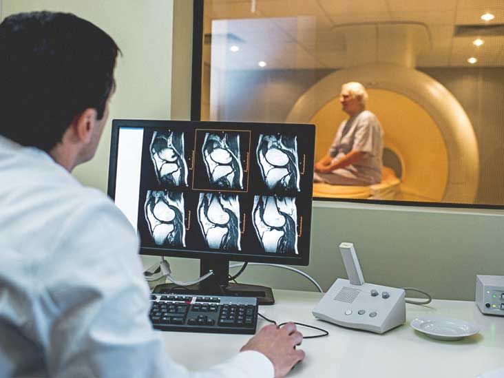
MRI (magnetic resonance imaging) uses strong magnets and radio waves to create “slices” of your bodyno ionizing radiation involved.[12] Think of it like a loaf of bread: each slice is an image. Stack enough slices and you get a detailed 3D view of your joint.
Musculoskeletal MRI is commonly used to evaluate joint disorders (including degenerative arthritis) and related structures like menisci, ligaments, and tendons, depending on the joint being scanned.[2] Different MRI sequences highlight different tissuessome make fluid light up, others make fat look bright, and some are designed specifically to spotlight inflammation.
What “arthritis” means on MRI: the big two patterns
While there are many types of arthritis, MRI findings often fall into two broad “themes”:
- Degenerative arthritis (most commonly osteoarthritis): more “wear-and-repair” changescartilage breakdown, bone remodeling, spurs.
- Inflammatory arthritis (like rheumatoid arthritis, psoriatic arthritis, some spondyloarthritides): more “immune-driven inflammation”synovitis, erosions, bone marrow inflammation, tendon/ligament insertion inflammation.
Real life is messier than categories. Osteoarthritis can have inflammatory features too (because joints are dramatic and enjoy overreacting). But this framework helps you interpret what you’re seeing.
What osteoarthritis looks like on MRI
Osteoarthritis (OA) is often described as cartilage gradually wearing down, which changes how the joint moves and how the bone responds.[9] X-rays are commonly used first, but MRI can reveal cartilage and soft-tissue changes in more detail, and it can also catch “bonus problems” that travel with OAlike meniscal tears or ligament degenerationespecially in the knee.[2]
1) Cartilage damage (the headliner)
Cartilage is the smooth “Teflon-like” layer that helps bones glide. On MRI, OA-related cartilage damage may be described as:
- Thinning (partial thickness loss)
- Fissuring (cracks/cleftslike a pothole starting to form)
- Full-thickness loss (bone surfaces are closer to rubbing, which can be painful)
In the knee, this often shows up in a specific compartment (medial, lateral, or patellofemoral), matching where your mechanics and loading are hardest on the joint. In the hip, it may show as cartilage loss and changes in the labrum (the ring of cartilage around the socket), depending on the clinical situation.
2) Bone spurs (osteophytes) and bone remodeling
Osteophytes are the body’s attempt to “stabilize” a stressed joint by building extra bonelike adding DIY brackets to a wobbling shelf.[9] MRI can show osteophytes, along with other subchondral (under-the-cartilage) changes such as:
- Subchondral sclerosis (bone becoming denser/harder)
- Subchondral cysts (fluid-like pockets sometimes called “geodes”)
3) Bone marrow lesions / “edema-like” signal
In OAespecially knee OAradiologists often comment on bone marrow lesions (BMLs), which appear as “edema-like” signal changes in the bone on fluid-sensitive MRI sequences.[7] These changes can fluctuate and sometimes correlate with pain, but they’re not a perfect one-to-one map: some people have impressive-looking MRIs with mild symptoms, and others have loud symptoms with subtle imaging.
4) Joint effusion and synovitis (yes, OA can do that)
OA isn’t always purely “wear and tear.” Many people have some degree of synovial irritation (synovitis) and extra joint fluid (effusion). MRI may show:
- Effusion: fluid collecting in the joint space
- Synovitis: thickening and inflammation of the joint lining (more obvious if contrast is used)
5) Meniscus and ligament “collateral damage”
Especially in the knee, OA commonly travels with meniscal degeneration/tears and sometimes meniscal extrusion (the meniscus shifting out of place), which can further increase stress on cartilage. MRI is a go-to tool for evaluating these related structures in context.[2]
What rheumatoid arthritis looks like on MRI
Rheumatoid arthritis (RA) is an inflammatory, immune-mediated arthritis. In early RA, X-rays may be normal even when inflammation is active, so MRI and ultrasound can be useful for early detection and assessing severity.[3] MRI can also help show the extent of active inflammation and early damage, including erosions that may be seen earlier than on X-ray.[5]
1) Synovitis (the classic RA feature)
RA often targets the synoviumthe lining of the joint. On MRI, synovitis can appear as synovial thickening and, if contrast is used, enhancement of the inflamed synovium. Clinically, this matters because persistent synovitis is part of what drives pain, swelling, stiffness, and long-term damage. Radiology case-based teaching from major orthopedic centers often describes RA as showing synovial proliferation/synovitis on MRI.[6]
2) Bone erosions (actual damage, not just “irritation”)
Erosions are areas where inflammation has “eaten into” bone near the joint. MRI can detect erosions, and these findings can appear earlier than on traditional radiographs in some patients.[5] The Arthritis Foundation also notes that imagingincluding MRIcan look for bone erosions in RA, which may start early and relate to future joint damage risk.[10]
3) Bone marrow edema / osteitis (a big red flag for progression)
In RA, MRI bone marrow edema (often interpreted as osteitisbone marrow inflammation) has been associated with later radiographic damage in research on early disease.[8] In practical terms: when clinicians see marrow edema plus synovitis in an RA pattern, they may interpret that as a sign of more aggressive inflammatory activity that deserves close attention.
4) Tenosynovitis and soft-tissue inflammation
RA doesn’t always stay politely inside the joint. It can inflame tendon sheaths (tenosynovitis), contribute to swelling around the joint, and affect nearby structures. MRI is helpful here because it shows tendons and soft tissues clearly, not just bone contours.
Other arthritis types: how MRI clues can differ
Psoriatic arthritis and “friends” (inflammatory arthritis with personality)
Psoriatic arthritis (PsA) can involve joints and also the entheseswhere tendons/ligaments attach to bone. Imagingincluding MRImay be used to evaluate joints, entheses, or spine involvement, depending on symptoms and suspected pattern.[11] (PsA can look different joint-to-joint, and sometimes one finger/toe is the “favorite” rather than a symmetrical pattern.)
Axial spondyloarthritis / ankylosing spondylitis (spine and SI joints)
For inflammatory back pain concerns, MRI can be used to evaluate the sacroiliac joints and spine for active inflammation (like marrow edema) and structural change. The key idea is similar: MRI can detect inflammatory changes earlier than plain radiographs in certain contexts.
Crystal arthritis (like gout): what MRI can and can’t do
MRI can show joint inflammation, erosions, and soft-tissue masses, but it isn’t always the best “crystal detector.” Diagnosis often relies on clinical pattern and, when needed, joint fluid analysis. MRI is more often used when the diagnosis is unclear or when clinicians are evaluating complications and the extent of tissue involvement.
Common phrases in MRI reports (and what they usually mean)
Here’s a translation cheat sheet for some of the most common arthritis-related MRI terms:
- Chondral thinning / cartilage loss: cartilage is wearing down
- Osteophytes: bone spurs (extra bone at joint margins)
- Subchondral sclerosis: bone beneath cartilage is getting denser
- Subchondral cysts / geodes: small fluid-like cavities in bone near the joint
- Effusion: extra joint fluid
- Synovitis / synovial hypertrophy: inflamed/thickened joint lining
- Bone marrow edema / BML: “edema-like” signal in bonecan relate to stress/inflammation depending on context[7]
- Erosions: bone damage near the joint, often emphasized in inflammatory arthritis patterns[10]
Why your doctor might order MRI for arthritis (and when they might not)
Many arthritis diagnoses start with a history, exam, and plain radiographs. But MRI is often considered when clinicians need information beyond what X-rays can show, such as:
- Early inflammatory arthritis suspicion: MRI/ultrasound can help identify early changes and severity when X-rays are still quiet.[3]
- Soft-tissue questions: cartilage, ligaments, tendons, menisci, and synovium are more visible on MRI than on X-ray.[1]
- Unclear diagnosis or unexpected symptoms: to look for alternate causes (stress fracture, osteonecrosis, tendon injury, etc.)
- Pre-surgical planning or targeted treatment decisions: especially when multiple structures are involved
Also: sometimes MRI is ordered because the story and exam suggest “arthritis plus something else,” and your clinician wants to make sure you’re not dealing with an additional issue that changes treatment.
What MRI can’t tell you (important, so you don’t panic-read your report)
MRI is detailedbut it’s not a mind reader.
- It can’t measure pain directly. Findings like effusion, synovitis, or BMLs may correlate with symptoms in some cases, but not perfectly.[7]
- It can’t always name the exact arthritis type. Many findings overlap. The pattern, distribution, clinical history, exam, and labs often do the final “diagnosis work.”
- It can show incidental age-related changes. Some cartilage wear or tendon degeneration can appear even in people without major symptoms.
- Timing matters. A flare today may look different than the same joint during a calmer week.
A realistic example: what arthritis might look like in two different joints
Example A: Knee osteoarthritis
A typical knee OA MRI might mention: cartilage thinning in the medial compartment, osteophytes, meniscal degeneration/tear, mild effusion, and a subchondral bone marrow lesion in the weight-bearing femur or tibia.[7] Functionally, this can translate to pain with stairs, standing from a chair, or long walksespecially if the meniscus is contributing to uneven load distribution.
Example B: Wrist/hand rheumatoid arthritis
An RA-pattern MRI might highlight synovitis, early erosions near the joint margins, and bone marrow edema/osteitis.[6][8] Clinically, this can line up with morning stiffness, swelling, tenderness across multiple joints, and symptoms that feel more “inflammatory” than purely mechanical. MRI and ultrasound may help in early-stage diagnosis and severity assessment when X-rays haven’t caught up yet.[3]
How to talk to your clinician about your MRI results
If your report feels like it was written for a secret club you were never invited to, here are questions that get useful answers:
- “Which findings explain my symptoms, and which are incidental?”
- “Does this look more degenerative (OA) or inflammatory (like RA/PsA)?”
- “Is there active inflammation (synovitis/edema) that changes treatment?”
- “What’s the next stepphysical therapy, medication, injections, lifestyle changes, further testing?”
- “Should we repeat imaging later, or track progress clinically?”
And a friendly reminder: don’t let an MRI report be your emotional support horoscope. It’s a tool, not a verdict.
Conclusion
On MRI, arthritis isn’t one single “look.” Osteoarthritis tends to show cartilage wear, bone remodeling, spurs, and sometimes bone marrow lesions and fluid. Inflammatory arthritis (like rheumatoid arthritis) often emphasizes synovitis, erosions, and bone marrow edema/osteitis patternssometimes earlier than X-rays can reveal.[3][5] The most helpful interpretation comes from combining MRI findings with your symptoms, exam, and (when needed) labsbecause your joint is part of your body, not a standalone mystery novel.
Medical note: This article is for education, not a diagnosis. If you have new swelling, severe pain, fever, rapidly worsening symptoms, or sudden inability to use a joint, seek prompt medical care.
Real-World Experiences: What It Feels Like to Get an Arthritis MRI
People often imagine an MRI as a dramatic, cinematic momentlike you’re being scanned by a sci-fi portal that will immediately reveal The Truth. In real life, it’s more like: “Please remove all metal, here are earplugs, try not to move, and yes, the machine will sound like a robot learning drum solos.” If you’re heading into an arthritis-related MRI, here are a few real-world experience patterns that come up a lot (names and details generalized, because your knee deserves privacy).
Experience 1: “I expected one picture. I got a whole photo album.”
Many patients are surprised by how long the scan takes. A knee MRI might feel quick, but if the technologist needs multiple sequences (or your joint is being extra uncooperative that day), it can take longer than you expected. The weird part is that you’re not “doing” anythingyour job is basically to become a statue with opinions. People often say the hardest part isn’t pain; it’s staying still when you want to adjust your leg by half an inch. A small tip that patients commonly find helpful: ask for cushions or supports early, before the scan starts, so you’re comfortable enough to hold the position. The moment the first loud sequence begins, you’ll understand why everyone becomes deeply grateful for ear protection.
Experience 2: “The report scared me. The doctor calmed me.”
A very common emotional arc goes like this: you read your report, see words like “degenerative changes,” “cartilage loss,” or “effusion,” and suddenly you’re Googling “how to replace an entire skeleton.” Then you talk to your clinician and realize: (1) some findings are common with age, (2) the report lists everything the radiologist sees, not just what’s causing symptoms, and (3) there are usually several reasonable next steps before anything dramatic. People often feel relief when the clinician points out which findings match their pain pattern and which ones are incidental. It’s also normal to feel frustrated if the MRI shows “a lot” but your symptoms are moderateor vice versa. Arthritis doesn’t always behave like a neat chart.
Experience 3: “I wanted a label. I got a plan.”
Some patients go into MRI hoping for a single, tidy diagnosis: “Is it osteoarthritis or not?” But many come out with something more useful: a targeted plan. For example, someone with knee pain may learn that cartilage wear is part of the story, but the biggest “action item” is a meniscus issue or a bone marrow lesion pattern that explains flare-ups with certain activities. Another person with suspected inflammatory arthritis may feel validated when imaging supports active inflammationbecause it changes how seriously a flare is treated and can guide earlier intervention. A recurring theme is that the most satisfying outcome isn’t a scary-sounding label; it’s clarity about what to do next (therapy, activity modification, medication, injections, or referrals).
Experience 4: “Small logistics made a big difference.”
People who report the best MRI experience often mention the same practical details: they told the technologist about claustrophobia up front, asked whether an open MRI was an option (when appropriate), requested a music channel if available, and confirmed whether contrast was expected. If contrast is used, patients often say the anticipation felt bigger than the actual injection. And almost everyone appreciates knowing the timeline for results beforehandbecause waiting can feel longer than the scan itself.
Bottom line: an arthritis MRI can be a powerful tool, but the human experience around it matters. Comfort, expectations, and a good post-scan conversation can turn a stressful “what’s wrong with me?” moment into a grounded “here’s what we can do” next step.