If you’ve ever read a radiology report and stumbled on the phrase “emphysematous changes”,
you’ve probably had the same reaction most humans do: “Is that… bad-bad, or just ‘don’t Google it’ bad?”
Let’s translate it into plain English.
Emphysematous changes usually means a scan (often a CT) shows areas of lung tissue that look like
emphysemadamage to the tiny air sacs (alveoli) where oxygen moves into your blood.
When those air sacs are injured, they can enlarge, merge, and lose their springiness, which makes it harder to push air out.
That “air-trapping” effect is a big reason people feel short of breath.
Important nuance: a report can mention “changes” even when symptoms are mildor when you don’t feel anything at all yet.
Think of it like a “check engine” light. It doesn’t tell you the whole story by itself, but it’s a clue worth taking seriously.
What “emphysematous changes” actually means
Emphysema is a form of chronic obstructive pulmonary disease (COPD).
COPD is an umbrella term that includes emphysema and chronic bronchitis, and it’s defined by airflow limitation that doesn’t fully go away.
Emphysema is specifically about structural damage: the walls between air sacs break down and the lung loses elastic recoil.
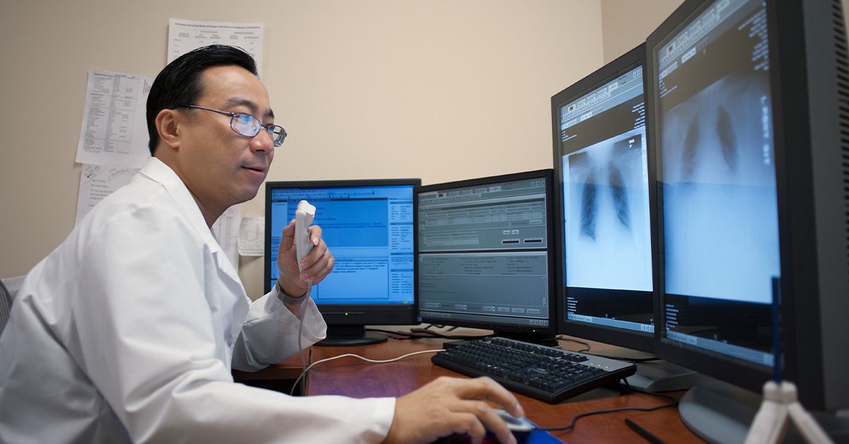
On imaging, radiologists may use “emphysematous changes” when they see patterns consistent with emphysemasuch as areas that look
darker (less dense), regions of overinflation, or larger air spaces (like blebs or bullae). A CT scan can detect emphysema earlier
and more clearly than a standard chest X-ray, so this phrase shows up a lot in CT reports.
Why it shows up on CT reports (and why that matters)
CT scans are excellent at spotting emphysema patterns and estimating how widespread they are. Sometimes the finding is discovered
incidentallylike during imaging for chest pain, a pre-op evaluation, or lung cancer screening.
Here’s the key point: a scan finding doesn’t equal a diagnosis by itself. The diagnosis and severity are usually confirmed with
pulmonary function tests, especially spirometry. Imaging shows structure; spirometry shows how well air moves in and out.
Many people have mild CT changes but near-normal breathing tests; others have significant symptoms with less dramatic imaging.
Your full picture comes from combining symptoms, test results, and risk factors.
Types of emphysema (the “patterns” behind the changes)
Emphysema isn’t one-size-fits-all. Doctors and radiologists often describe emphysema by where in the lung unit it starts and
where it tends to show up in the lungs. These patterns matter because they hint at causes and risks.
1) Centrilobular (centriacinar) emphysema
This is the most common type. It tends to affect the central portion of the lung lobule and is strongly linked with
cigarette smoking. It often shows up more in the upper lobes.
Example: A long-time smoker gets a CT for a persistent cough. The report says “mild centrilobular emphysematous changes, upper-lobe predominant.”
That pattern fits smoking-related injury and can be an early warning signespecially if spirometry is starting to show obstruction.
2) Panlobular (panacinar) emphysema
Panlobular emphysema affects the lobule more uniformly and is classically associated with alpha-1 antitrypsin deficiency (AATD),
a genetic condition that reduces protection against enzymes that can damage lung tissue. It’s often more noticeable in the lower lobes.
Example: A person in their 30s or 40sespecially with limited smoking historyhas significant shortness of breath.
Their CT suggests lower-lobe predominant panlobular changes. That’s a scenario where clinicians often consider testing for AATD.
3) Paraseptal (distal acinar) emphysema
Paraseptal emphysema appears near the lung edgesclose to the pleura (the lining) or along fissures.
It can form small air pockets called blebs or larger ones called bullae.
Some people have this pattern without major airflow obstruction, but it can be associated with a risk of a
spontaneous pneumothorax (collapsed lung) if a bleb ruptures.
Example: A tall young adult with sudden sharp chest pain and shortness of breath is found to have a pneumothorax.
Imaging later notes paraseptal emphysema with blebs near the lung apex. Not everyone fits this storybut it’s a classic one.
4) Irregular (paracicatricial) emphysema
This pattern is associated with scarring (fibrosis) or prior lung injury. It’s “irregular” because it doesn’t follow the tidy lobular patterns.
You might see it near old infections, prior inflammation, or other lung diseases that leave scarred areas behind.
Common causes and risk factors
Emphysematous changes usually result from long-term exposure or vulnerability that gradually damages lung tissue.
The most common driver is tobacco smoke, but it’s not the only one.
Major risk factors include
- Smoking (including long-term heavy exposure).
- Secondhand smoke and chronic exposure to irritants.
- Workplace exposures (dusts, fumes, chemicals) and environmental air pollution.
- Genetic risk, especially alpha-1 antitrypsin deficiency.
- History of asthma or repeated lung infections (risk can compound over time).
Also worth noting: some people with COPD/emphysema have never smoked. If your report shows emphysematous changes and you’re a non-smoker,
your clinician may think more carefully about other exposures, asthma history, and genetic testing when appropriate.
Symptoms: what you might notice (or not notice yet)
Early emphysematous changes can be sneaky. You might feel completely fineespecially if the changes are mild and your lungs have “reserve.”
As the condition progresses, symptoms tend to show up gradually.
Common symptoms
- Shortness of breath, especially with exertion (stairs become personal enemies).
- Chronic cough and/or mucus (more common when chronic bronchitis overlaps).
- Wheezing or chest tightness.
- Reduced exercise tolerance, fatigue, or needing more time to recover after activity.
- Unintended weight loss in more advanced disease (breathing becomes “expensive” work).
A helpful clue is a shift in your “baseline”: you used to walk the parking lot easily, and now you’re planning your route around benches like you’re on a scenic tour.
That change matterseven if it happens slowly.
How clinicians confirm the diagnosis and assess severity
1) History and exam
Clinicians look at symptoms, smoking history, occupational exposures, family history, and whether you’ve had frequent bronchitis or pneumonia.
A physical exam may be normal early on, which is why testing matters.
2) Spirometry (the cornerstone test)
Spirometry measures how much air you can blow out and how fast you can blow it out. It helps determine whether airflow is obstructed.
It’s one of the main tests used to diagnose COPD and gauge its severity.
3) Other pulmonary function tests
Clinicians sometimes add tests like diffusing capacity (DLCO) (often lower in emphysema) or lung volumes (to assess hyperinflation and air trapping).
These tests help separate “airway” problems from “air sac” problems and can guide treatment decisions.
4) Imaging: chest X-ray vs CT
Chest X-rays can sometimes show signs of hyperinflation, but CT is better at detecting emphysema early and describing the pattern.
CT findings may also help identify large bullae or determine whether someone might be evaluated for specific procedures in advanced cases.
5) Testing for alpha-1 antitrypsin deficiency (when appropriate)
AATD testing may be considered if emphysema appears at a young age, seems severe compared with smoking history, runs in the family,
or shows a pattern suggestive of panlobular/lower-lobe disease.
Possible complications (the things we’re trying to prevent)
Emphysema can increase the risk of complications, especially as it becomes more advanced or overlaps with chronic bronchitis.
These can include:
- Exacerbations (flare-ups) often triggered by infections or irritants.
- Respiratory infections such as pneumonia.
- Low oxygen levels in more severe disease.
- Pneumothorax (collapsed lung), particularly with blebs/bullae.
- Pulmonary hypertension and strain on the right side of the heart in advanced cases.
Treatment and management: what actually helps
There’s no magic “undo” button for destroyed alveolar walls. But there are many ways to reduce symptoms, improve function,
and slow progression. Treatment is usually personalized based on symptoms, spirometry results, exacerbation history, and imaging pattern.
1) Quit smoking (the single biggest win)
If smoking is part of the picture, quitting is the most effective step to slow further lung damage.
It’s not a moral lecture; it’s a physics problem. Remove the irritant, and the injury rate drops.
2) Medications
Many people benefit from inhaled medications that help open the airways (bronchodilators). In some casesparticularly with frequent exacerbations
clinicians may add inhaled anti-inflammatory medicines. The exact medication plan depends on your symptoms and test results.
3) Vaccines and prevention
Staying up to date on recommended vaccines (like influenza and pneumococcal vaccines) can reduce the risk of infections that trigger flare-ups.
Avoiding indoor smoke, improving ventilation, and using protective gear at work (when applicable) also matter.
4) Pulmonary rehabilitation (secretly one of the best tools)
Pulmonary rehab is a structured program that combines supervised exercise, breathing techniques, education, and support.
It helps people breathe better, improve stamina, and feel more confident doing daily activities.
If you picture it as “gym class for lungs,” you’re not wrongexcept this gym class actually makes sense.
5) Oxygen therapy (for those who need it)
If oxygen levels are low, supplemental oxygen may be prescribed to reduce strain on the body and improve quality of life.
Oxygen is a treatment for low oxygennot a sign you “failed.” It’s simply the right tool for the job when needed.
6) Procedures and surgery (selected cases)
In advanced emphysema, especially when certain lung areas are far more damaged than others, some people may be evaluated for interventions such as:
- Lung volume reduction surgery (LVRS) (removing the most damaged portions to help the remaining lung work more efficiently).
- Bullectomy (removal of a very large bulla that compresses healthier lung).
- Bronchoscopic lung volume reduction (such as endobronchial valves in carefully selected candidates).
- Lung transplant (for severe disease in select individuals).
These options require specialized evaluation and aren’t for everyone, but they’re important to know aboutbecause “nothing can be done” is often untrue.
Living with emphysematous changes: practical, real-world tips
Track your baseline
A simple habit: notice how far you can walk comfortably, how many stairs you can do, and how often you reach for rescue inhalers (if prescribed).
Changes over weeks to months are useful data for your clinician.
Use breath-saving strategies
Techniques like pursed-lip breathing can help empty trapped air and reduce the “I can’t get a full breath” feeling.
Pulmonary rehab teaches these skills in a way that’s actually usable outside a textbook.
Make your environment less annoying to your lungs
Smoke, strong fragrances, dust, and poor air quality can trigger symptoms. Think of it as reducing pop-up ads for your airways.
Air filters, better ventilation, and avoiding indoor burning (like smoking indoors) can help.
When to seek urgent care
Seek urgent medical attention if you have severe or rapidly worsening shortness of breath, chest pain, bluish lips or face,
confusion, fainting, or symptoms that feel dramatically different from your normal baselineespecially if you suspect a pneumothorax
(sudden sharp chest pain with shortness of breath).
Frequently asked questions
Is “emphysematous changes” the same as emphysema?
It usually means the scan shows features consistent with emphysema. Whether it meets clinical criteria and how significant it is
often depends on symptoms and pulmonary function testing.
Can emphysema be reversed?
Structural damage to alveolar walls is generally not reversible, but symptoms and day-to-day function can improve significantly with treatment,
rehab, and risk-factor control (especially smoking cessation).
Can you have emphysema if you never smoked?
Yes. While smoking is the most common cause, emphysema/COPD can also be related to secondhand smoke, occupational exposures,
asthma history, air pollution, and genetic factors like alpha-1 antitrypsin deficiency.
Experiences people commonly describe (and what they often learn the hard way)
People hear “emphysematous changes” and imagine their lungs turning into Swiss cheese overnight. In real life, it’s usually more like a slow
negotiation between your body, your habits, and gravityespecially gravity. One of the most common stories goes like this:
“I didn’t feel sick. I just… got slower.” The first clue may be that you’re taking the elevator more often or avoiding the back aisle at the grocery store
because it’s a whole expedition. You may not call it shortness of breath; you call it “being out of shape.” But when the pattern repeatsstairs,
brisk walking, carrying laundrypeople begin to notice a theme.
Another classic experience is the emotional roller coaster of reading the report. Many people go from “It’s probably nothing” to “I definitely have
five minutes to live” in the time it takes a browser to load. The helpful reframe: emphysematous changes are a signal, not a sentence.
Clinicians often explain that imaging and spirometry are like two camera angles on the same story. The report might show mild changes, and your
breathing tests may be reassuringmeaning the focus becomes prevention and protecting what you’ve got. Or spirometry may reveal more impact than
expected, which helps target treatment sooner rather than later.
People who start pulmonary rehab often describe it as the first time someone taught them how to breathe with intention. It’s not just treadmill time.
It’s learning why you get that “air hunger” feeling, how pacing reduces panic, and how pursed-lip breathing can make exhaling more effective when air
gets trapped. Many participants joke that rehab is like joining a club where everyone understands the weird things you do to conserve breathlike
strategically pretending to admire a painting in the hallway when you’re actually just recovering.
Quitting smokingwhen it appliesoften comes with its own set of stories. People describe it as breaking up with someone who was bad for them but
“always there,” which is both funny and painfully accurate. What stands out is how often the goal shifts from “I must never cough again” to
“I want to walk with my kid/grandkid without planning a rest stop.” Clinicians emphasize that quitting doesn’t regrow damaged air sacs, but it can
slow further injury. Many people say their cough improves and their stamina stops declining as quicklysmall wins that add up.
Those with bullae or paraseptal changes sometimes live with a low-level worry about a collapsed lung. The good news is that most days are normal.
The practical takeaway is knowing what “not normal” feels like: sudden sharp chest pain and abrupt breathlessness should be checked right away.
Having a plan reduces fear. It turns “What if?” into “If X happens, I do Y.”
Finally, people with alpha-1 antitrypsin deficiency often describe a different journey: confusion about why lung disease showed up “too early,” relief at
having an explanation, and a new focus on family screening and avoiding exposures. Their experience highlights a big theme in emphysematous changes:
the earlier you understand the pattern and the cause, the more options you havewhether that’s targeted treatment, workplace changes, or simply getting
serious about prevention while the lungs still have plenty of reserve.
Conclusion
“Emphysematous changes” is medical shorthand for imaging features that suggest emphysema-related lung damage. The phrase can represent anything from
early, mild findings with no symptoms to more advanced diseaseso the next step is usually context: symptoms, exposures, and lung function tests.
With the right planrisk reduction, medications when needed, pulmonary rehab, and monitoringmany people breathe better, stay active longer, and keep
life bigger than their diagnosis.